There is no special significance to the "crown" of the eukaryotes, it is merely the point where we don't know for sure what the exact relationships are so we just put all of the major branches together here until we find out the true phylogeny. This in no way implies that these lineages arose simulataneously. Since there is little phylogenetic structure to show here, I will just enumerate the groups in a list and you will need to be aware that they can be in any order.
The major groups described here are the RHODOPHYTA, STRAMENOPILES, ALVEOLATES, FUNGI & AMOEBAS.
RED ALGAE (RHODOPHYTA)
STRAMENOPILES
The STRAMENOPILES includes the brown algae, the golden algae
(diatoms) and the water molds. The group is well supported by DNA evidence
although it is very diverse morphologically. The three parted , hollow
hairs usually found on the tinsel flagellum of the two flagellae common
in gametes or spores of the group is the only morphological synapomorphy
for the group and it has been lost in the diatoms.
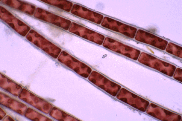
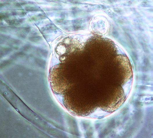
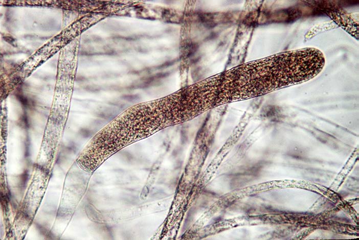
This group includes the Water Molds (OOMYCOTA). Shown in the next two photos is the genus Saprolegnia, the first is a sporangium of nonmotile female gametes (fertilized) and the second is of a sporangium of asexually derived flagellated spores. The water mold Phytophorawas responsible for the great potato famine in Ireland. These were once considered fungi although the only thing that they really share with fungi is a lack of chlorophyll.
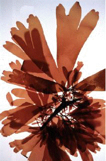
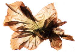
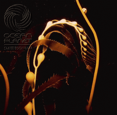
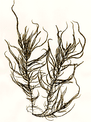
The Stramenopiles also include the BROWN ALGAE (PHAEOPHYTA) which ranges
from tiny filamentous microscopic organisms to some of the largest organisms
on earth (the great sea kelps) as well as the familiar Fucus and
Sargassum
that often wash up on Florida beaches. Many have highly developed body
plans which include distinct stems, holdfasts and air bladders. The brown
algae are widely used medicinally as well as for Parkelp (a food additive)
and Kombu & Wakame (used in oriental cooking). They are also the source
of Algin which is ubiquitous as a thickener and smoothing agent in jellies,
ice cream etc.
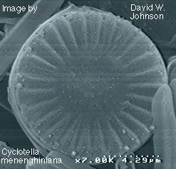
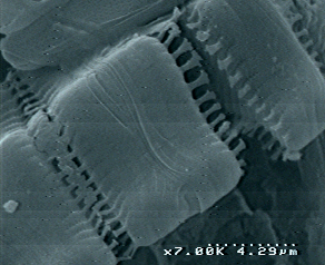
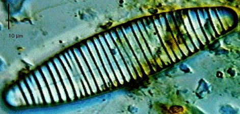
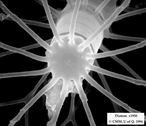
The third major group that we will look at in the Stramenopiles is the group made up of the golden alga / the diatoms , collectively the CHRYSOPHYTA. They may be the most beautiful of all the groups often having a silica (glass) cell wall in 2 parts that fit together like a Petrie plate. They have chlorophyll a & c.
This group includes the Dinoflagellates, Ciliates & Sporozoans.
The major synapomorphy for this group is the peculiar type of body openings that they have called alveoli.
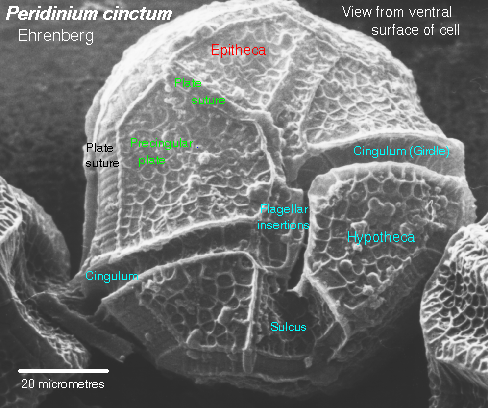
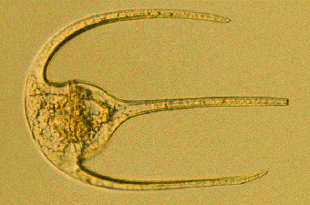
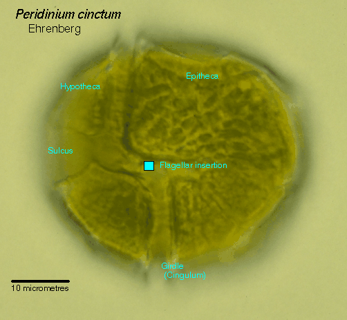
Dinoflagellates: These are plantonic, free floating, single celled organisms generally with their outer body covered with large, sometimes ornamented, cellulose plates with two furrows, one going wholly around the body. They have 2 flagellae, one that sits in the longer body furrow while the other is perpendicular to it, partially in the other furrow but extending out from it. These are the organisms that cause red tide, many are poisonous to fish. They are autotrophic and their chloroplast contains chlorophyll a & c and is more autonomous than those of other groups. There are no spindle fibers formed in mitosis and meiosis because the chromosomes are attached to the nuclear membrane. The big ones shown here are Peridinium (from the Protist Image Database), the smaller ones in the middle are other genera from the Tsukuba Univ. Server.

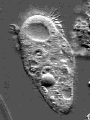
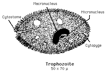
Ciliates: These are single celled organisms with a high degree of complexity. They are covered with cilia and are heterotrophic. They have both a macronucleus and a micronucleus, contractile vacuole, food vacuoles etc. The Paramecium on the BSC2005 web page (from Cells Alive! http://www.comet.net/quill/Index1.htm) is a ciliate.
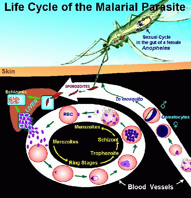
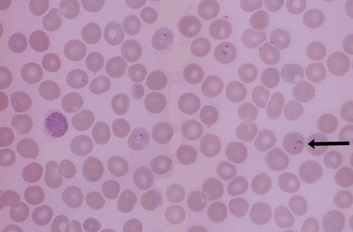
AMPICOMPLEXANS (Sporozoans): These are the final group of alveolates that we will look at. They are all parasites and normally have very complex life cycles. The genus toxoplasma is a major cause of birth defects in humans and livestock, but the one that we will consider for our example is Plasmodium, the organism that causes malaria. This disease kills more than 1 million people a year (many children) and has debilitated countless others (including your instructor). Plasmodium vivax and P. falciparum are the primary agents for malaria. Humans are infected by a mosquito with the sporozoites which then undergo mitosis in the liver and mass release into the red blood cells where they undergo mitosis again then mass release again and undergo meiosis into the blood which the mosquito sucks up; in the mosquito the gametes form a zygote, go into a cyst stage and then transform into the active sporozoite stage to infect another human.
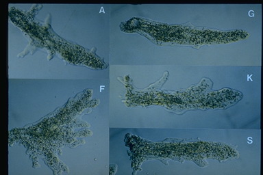
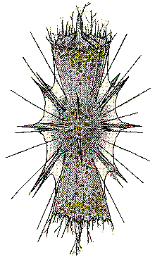
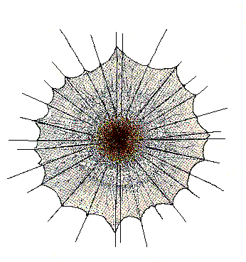
Another group - the radiolarians have modified pseudopods that stick out in all directions. These are from Schewiakoff 1926 - an early work on the group.